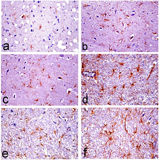
Fig 12. Glial fibrillary acidic protein (GFAP)-immune stained astrocytes in brain sections of rats.
The micrographs represent the following groups: (a) Vehicle, (b) Met, (c) Saxa, (d) D-gal, (e) D-gal/Met, and (f) D-gal/Saxa. (GFAP immunohistochemical stain, X40). Vehicle, rats treated with distilled water (1.5 ml/kg/day, s.c); Met, rats treated with metformin (500 mg/kg/day, p.o); Saxa, rats treated with saxagliptin (1 mg/kg/day, p.o); D-gal, rats treated with D-galactose (150 mg/kg/day, s.c); D-gal/Met, rats treated with D-galactose and metformin; D-gal/Saxa, rats treated with D-galactose and saxagliptin.

