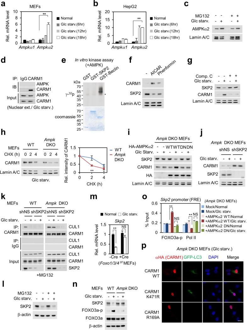
Extended Data Figure 5. AMPKα2 accumulates in the nucleus leading to repression of SKP2 and stabilization of CARM1 under nutrient-starved conditions.
a, b, qRT–PCR of Ampka1 and Ampka2 in MEFs (a) and HepG2 cells (b) upon glucose starvation. c, The nuclear AMPKα2 expression level was analysed in the absence or presence of MG132. d, Binding between CARM1 and AMPK was assessed. e, In vitro kinase assay with AMPK. f, MEFs were treated with AICAR (1 mM) or phenformin (2 mM) for 4 h. The nuclear fraction was analysed by immunoblot. g, MEFs were deprived of glucose in the absence or presence of 10 µM compound C and the nuclear fraction was analysed by immunoblot. h, Left, cycloheximide treatment in wild-type and Ampk DKO MEFs. Right, protein half-life of CARM1 was quantitatively defined. i, j, Ampk DKO MEF lysates were analysed by immunoblot. k, CARM1– CUL1 interaction was analysed after SKP2 knockdown in wild-type and Ampk DKO MEFs. l, SKP2 expression levels were analysed in the absence or presence of MG132. m, Foxo1/3/4f/f MEFs infected with Cre virus were analysed for Skp2 mRNA. n, SKP2 and phosphorylated FOXO3a were analysed by immunoblot. o, ChIP assay of the Skp2 promoter. Data are mean ± s.e.m.; n = 3. *P < 0.05, **P < 0.01 (one-tailed t-test) (a, b, h, m, o). p, Representative confocal images. Scale bar, 20 µm.

