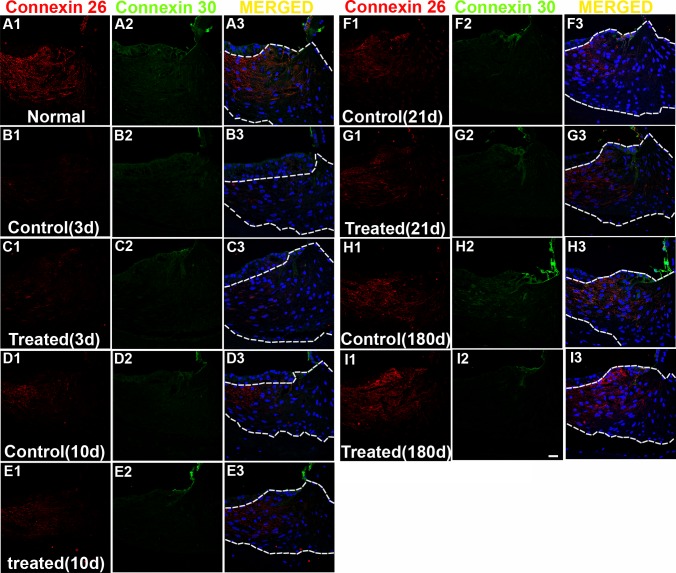
Fig 13. Representative confocal images of connexin 26 (red) and 30 (green) labeling collected from the spiral ligament.
Nuclei of cells were stained by DAPI (blue). Merged images are shown in the column 3 (A3-I3, merged). Positive Connexin 26 labeling (red) is observed in fibrocytes in the spiral ligament of normal (naïve) controls (A1). Decreased connexin 26 labeling (column 1) is observed in the spiral ligament of chinchilla all time points after noise exposure (B-H) except in the treated group at 6 months after noise exposure(I). No or very low CX-30 labeling is observed in fibrocytes of the spiral ligament of chinchilla while the root cells show positive Cx-30 labeling (A2-I2). Scale bar = 20 μm in I3 for A1-I3.

