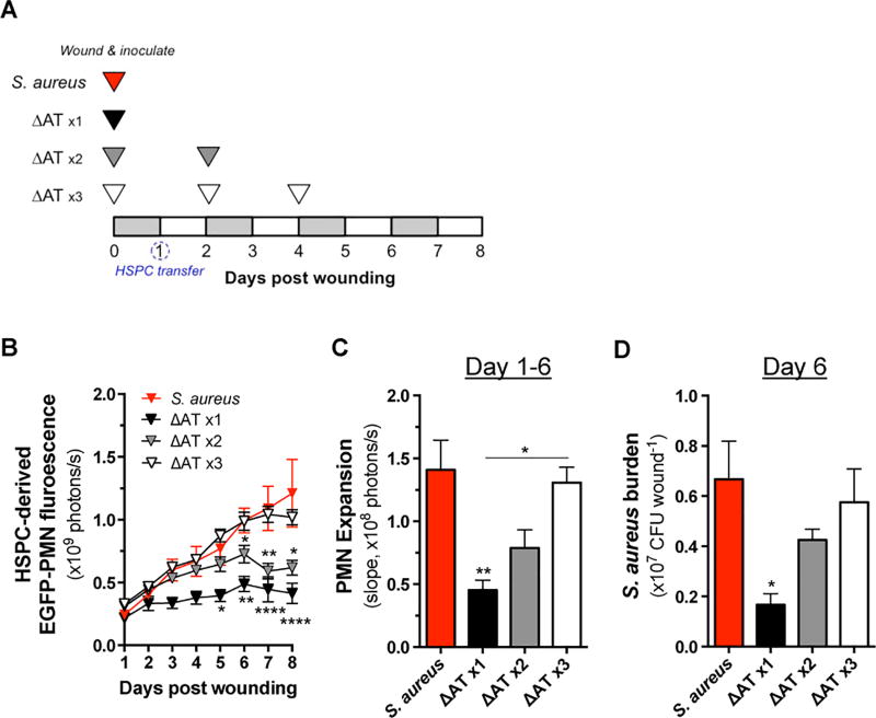
Figure 4. HSPC expansion within the wound is responsive to bacterial abundance.
(A) C57BL/6 mice were wounded and inoculated with 1×10⁷ CFU of S. aureus or ΔAT. At 24 hours post wounding, mice received a local injection of 5×10⁵ HSPC from LysM-EGFP donor mice. Some animals that received ΔAT at day 0 received additional inoculations of equivalent CFU at day 2 or days 2 and 4 post wounding as indicated. (B) EGFP-PMN fluorescence at wound sites was quantified daily. (C) The rate of increasing EGFP-PMN fluorescence was determined based on the slopes depicted in panel B. (D) Bacterial burden from wounds of mice from each experimental group was determined from homogenized wound tissue collected at day 6 post wounding. Data represent 4–10 mice per group and are expressed as mean±SEM. * p < .05, ** p < .01, versus S. aureus or as indicated. 1-way ANOVA (p < .01 and p < .01 for 4B and 4C, respectively) with Tukey’s multiple comparisons test. 1-way ANOVA (4D, ns) with Dunnett’s multiple comparisons test.

