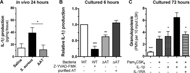
Figure 6. IL-1β and TLR2 activation signals granulopoiesis of bone marrow derived HSPC.
(A) IL-1β protein expression from homogenized wound tissue collected 24 hours post wounding and inoculation with saline or S. aureus versus ΔAT as measured by ELISA. (B) Bone marrow derived PMN were stimulated with live S. aureus or ΔAT in the presence and absence of a caspase1 inhibitor (Z-YVAD-FMK) and purified alpha-toxin. IL-1β protein levels were measured in culture supernatants by ELISA. Non-stimulated cells showed protein levels below the limit of detection for the ELISA (data not shown). Data normalized for each mouse to IL-1β produced by WT S. aureus. (C) In vitro expansion of LSK from BM-derived HSPC cultured for 72 hours with Pam3CSK4 alone, IL-1β alone, Pam3CSK4 + IL-1β, Pam3CSK4 + IL-1β + IL-1RA, or vehicle control. Data are expressed as mean±SEM and represent at least 3 independent experiments. * p < .05, ** p < .01, *** p < .001, WT + Z-YVAD-FMK, ΔAT, or ΔAT + AT versus WT alone (B) or vehicle, Pam3CSK4 alone, IL-1β alone, or Pam3CSK4 + IL-1β + IL-1RA versus Pam3CSK4 + IL-1β (C). 6A: two-tailed unpaired t-test; 6B-C: 1-way ANOVA (p<0.0001 for 6B and 6C) with Tukey’s multiple comparisons test.

