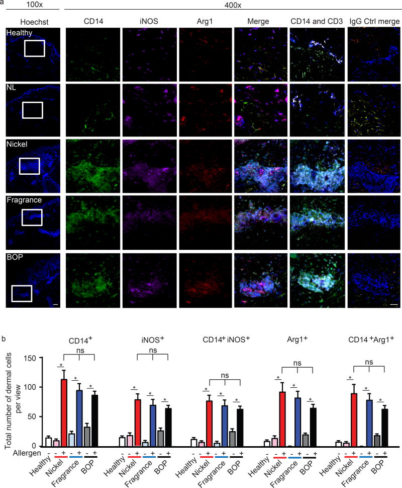
Figure 1. iNOS and Arg1-expressing MΦ infiltrate ACD lesions.
(a) Representative immunohistochemistry analysis depicting CD14 (green), iNOS (purple), Arg1 (red), CD3 (white), Hoechst (blue) expression in ACD skin (Nickle, Balsam of Peru; BOP, and fragrance), non-lesional (NL), and healthy controls. Data are representative of 1–2 patient samples per tested condition with similar results, original magnification ×100(left) and x400 with scale bar 100 µm and 50 µm, respectively.
(b) Analysis depicting total numbers of dermal CD14+, iNOS+, Arg1 and co-expression of CD14 with iNOS and Arg1 in ACD lesions (Nickel, Balsam of Peru; BOP, and fragrance), non-lesional (NL), and healthy samples. Data are expressed as positive cells ± SEM from at least three seperate microscopic fields, 400x, from 1–2 patients, *p<0.05.

