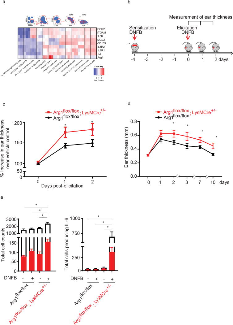
Figure 3. Arg1 deletion in mono/MΦ promotes CHS in vivo.
(a) Heat map showing various genes which were 2- log fold up or down regulated in murine monocyte, macrophage and DC populations under steady state or following DNFB (microarray GSE49358 from Tamoutounour et al., 2013 (18)) Normalized gene expression was plotted across the samples after z-score normalization. Genes are clustered using a correlation distance with complete linkage.
(b) A timeline indicates experiment chronology.
(c) Arg1flox/flox; LysMCre+/−and Arg1flox/flox were sensitized and elicited with DNFB. Ear swelling was measured daily and is depicted as mean increase over vehicle control ± SEM from 15–22 mice, *p<0.05
(d) Arg1flox/flox; LysMCre+/−and Arg1flox/flox mice were sensitized for 5 times before re-elicitation with DNFB on the ear. Ear swelling was measured daily and is depicted as mean ± SEM from 5 mice, *p<0.05, data were analyzed by two-way ANOVA test followed by least-significant differences multi-comparison (LSD) test.
(e) Left: Total number of CD45+ cells (white bar) and CD45+CD11b+CD64+ (red bar ), and CD45+CD11b+CD64+( red bar ); Right : Total number of CD45+ cells producing IL-6 (white bar) and CD45+CD11b+CD64+ producing IL-6 (red bar) in skin from Arg1flox/flox; LysMCre+/−and Arg1flox/flox mice at 12 hrs post-DNFB. Data are summarized as mean ± SEM from 3 mice, *p<0.05

