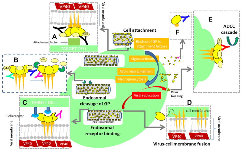
Figure 5.
Known anti-GP mAbs interfere with key GP functions at different stages of the progression of EBOV infection. (A) Upon interaction with the host cell through attachment factors (not precisely receptors), a complex series of biochemical signals are triggered, eventually leading to EBOV entry through macropinocytosis and endosome formation. Several mAbs are known to interfere with virus-cell attachment (black mAb). (B) Transmembrane GP is cleaved by proteases (dark green symbols) within endosomes. This enzymatic cleavage removes the MLD region (indicated in a lighter shade of yellow) and the glycan cap exposing the RBD at GP1. Several mAbs bind cleaved forms of GP and, by doing so, interfere with GP binding to cell receptors (pink antibody) and (C) further enabling the interaction of viral GP with cell receptors (blue ovals) through the RBD. The interaction of cleaved GP with cell receptors (v.gr. NCP1) triggers virus-cell membrane fusion. After GP binding to receptors, (D) GP is further cleaved, and a significant portion of GP1 is lost, the remaining GP1-GP2 peptide undergoes a geometrical rearrangement to initiate fusion. Some antibodies simultaneously bind epitopes at G1 and G2 interfering with the series of structural arrangements required for virus-cell membrane fusion. (E) Anti-GP mAbs may also bind to the GP molecules exposed at the surface of infected cells, marking them for further host immune response and attacking through mechanisms including antibody-dependent cell-mediated cytotoxicity (ADCC). (F) Binding to sGP conceivably decreases the number of mAb units available to interfere with transmembrane or cleaved GP.

