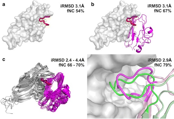
Fig. 2.

Visualization of the structures resulting from each of the modeling steps of the EphB4-EphrinB2 protein–protein interaction. For each modeling step interface RMSD (iRMSD) and fraction of native contacts (fNC) are provided. a Result of SLiM (from EphrinB2) docking to the EphB4 receptor (the SLiM is marked in red, EphB4 is visualized as a gray surface), b superimposition of the EphrinB2 structure on the docked SLiM peptide (the SLiM is marked in red, EphB4 is visualized as a gray surface, the structure of EphrinB2 is colored in magenta), c results of the complex refinement. The left panel shows the set of 10 models from the GalaxyRefine procedure. The right panel focuses on the interaction interface of the EphrinB2 SLiM (magenta) and EphB4 (gray surface) obtained from the FG-MD refinement procedure and its comparison with the experimental complex structure (PDB ID: 2HLE, shown in green)
