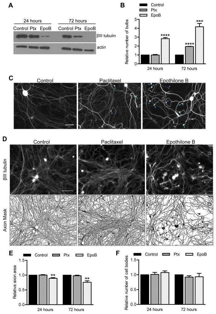
Fig 2. The neurotoxicity of epothilone B and paclitaxel correlates with their microtubule stabilization.
Cultured DRG neurons were treated with 2 nM paclitaxel or epothilone B starting at 3 DIV and treatment was refreshed every 8 hr for 24 or 72 hr.
A) After epothilone B treatment, lower levels of soluble tubulin remained than after paclitaxel due to the greater potency of epothilone at stabilizing polymerized tubulin. Actin serves as a loading control.
B) Quantification of retraction bulbs per field. The number of retraction bulbs in control cultures was set as 1 and fold changes in response to paclitaxel and epothilone B treatment were calculated. ***p<0.001, ****p<0.0001; Student’s t test.
C) Representative fields immunostained for βIII tubulin with retraction bulbs (blue arrowheads) at 72 hr, as quantified in (B). Scale bar, 50 μm.
D–E) To assess axon area after 72 hr of treatment, cell bodies were removed from the images and an axon mask was created. Axon area was then quantified and expressed as fold change compared control. Scale bar, 100 μm. **p<0.01, Student’s t test.
F) The number of cell bodies per field was counted from images like those in (D) and expressed as fold change compared to control. p>0.05, Student’s t test.
For (A–F), n=4 experiments. Ptx, paclitaxel.

