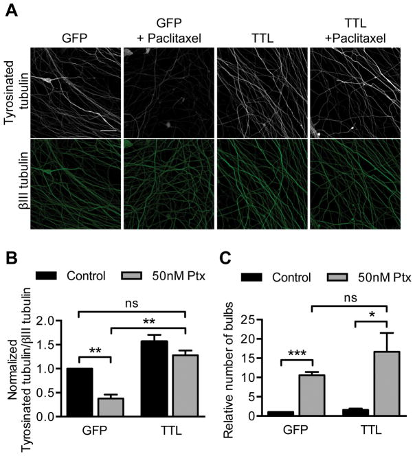
Fig 5. Maintaining control levels of tyrosinated tubulin after paclitaxel treatment does not rescue paclitaxel neurotoxicity.
TTL or GFP control lentivirus was added to DRG neurons at 0 DIV and treated with 50 nM paclitaxel for 48 hr starting at 5 DIV prior to immunostaining for tyrosinated tubulin and βIII tubulin at 7 DIV.
A–B) The fluorescence intensity of tyrosinated tubulin signal from axons was normalized to βIII tubulin signal, and fold changes from control were calculated. Scale bar, 20 μm. **p<0.01, ns p=0.18; Student’s t test.
C) Number of retraction bulbs per field was quantified and is expressed as a fold change relative to control. ***p<0.001, *p=0.037, ns p=0.28; Student’s t test.
For (A–C) n= 3 experiments. Ptx, paclitaxel.

