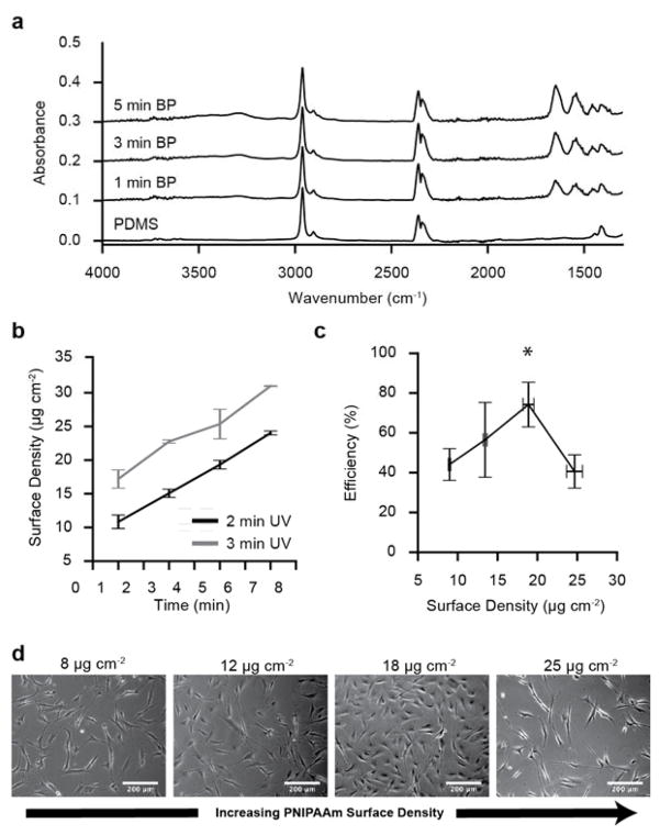
Figure 3.
Fourier transformed infrared spectra and cell attachment efficiency of PNIPAAm-PDMS substrates. (a) FT-IR absorbance spectra for PNIPAAm-PDMS substrates soaked in BP (acetone) for 0–5 min and then grafted with 3 min UV exposure. (b) Mean PNIPAAm surface density (± s.e.m.) for substrates soaked in BP for varying durations (1–7 min) and then PNIPAAm grafted with 2 or 3 min UV exposure (N=5). (c) Mean vascular smooth muscle cell seeding efficiency (± s.d.) for PNIPAAm-PDMS substrates with varying PNIPAAm surface density (N=3). (d) Phase contrast microscopy representative images of vascular smooth muscle cell attachment on PNIPAAm-PDMS with different graft densities taken 18 hours after seeding.

