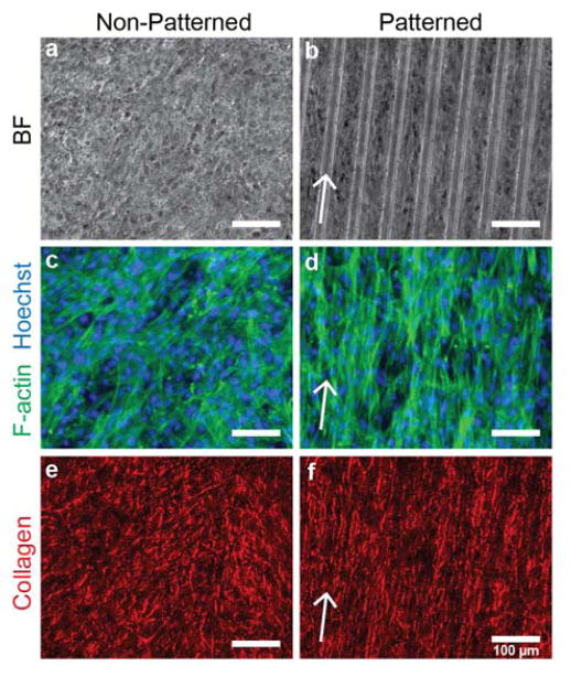
Figure 5.
Vascular smooth muscle cell sheet structure on non-patterned and patterned substrates. (a,b) Phase contrast images of vascular smooth muscle cell sheets grown on non-patterned and patterned substrates for 17 days. (c,d) F-actin structure for non-patterned and patterned VSMC sheets (green). Cell nuclei are labeled with Hoechst (blue). (e,f) Collagen structure (red) of VSMC sheets after picrosirius red stain. Arrows correspond to direction of micro-pattern. Scale bar indicates 100 μm.

