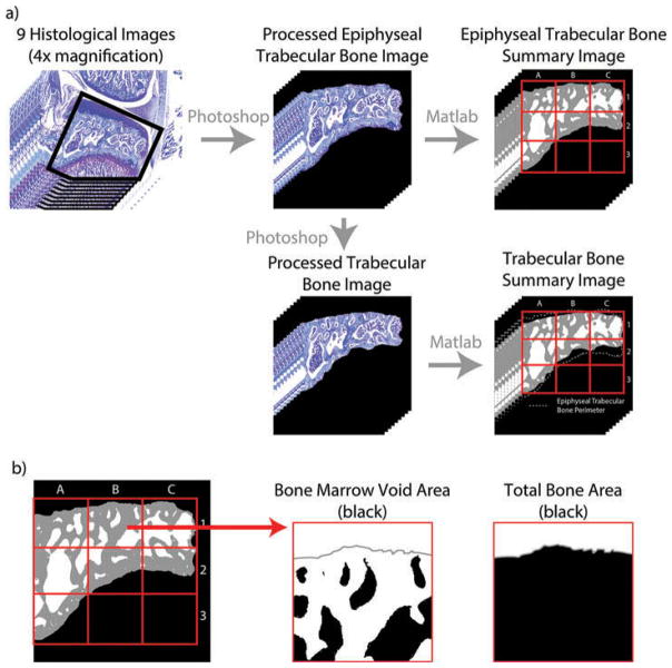
Figure 2.
Calculating trabecular bone area. a) The image is first prepared in Adobe Photoshop by rotating the image such that the tibial plateau is horizontal and erasing any points where the dehydrated bone marrow is contiguous with the trabecular bone. To prepare the epiphyseal trabecular bone images, surrounding tissue is then removed leaving the subchondral bone area between the articular cartilage and epiphyseal plane. From these images, the trabecular bone images are created by removing the bone between the articular cartilage and bone marrow voids and between the epiphyseal plane and bone marrow voids. b) This Photoshop prepared image is then loaded into a custom MATLAB script, where trabecular bone area is calculated over the entire subchondral bone area and within 9 pre-defined zones (red grid).

