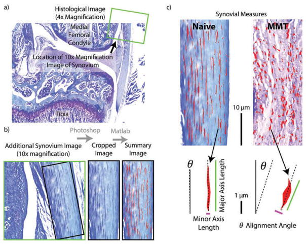
Figure 3.
Calculating synovium measures. a) The 10x magnification synovial image (green square) is taken proximal to the meniscus in the medial compartment. b) As in Figure 2, the synovial image is first prepared in Adobe Photoshop by rotating the image to vertical and cropping the image such at only synovial tissue is included. c) This Photoshop prepared image is then loaded into a custom MATLAB script, where subintimal cells are identified and measures of cellular density and morphology are calculated.

