Figure 6.
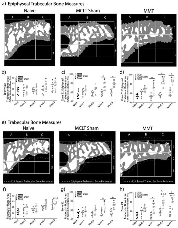
Trabecular Bone Area. a) Representative images of processed epiphyseal trabecular bone area bone (ETBA) images for naïve, MCLT sham, and MMT groups. b) ETBA increases in MMT animals by week 4 post-surgery and in MCLT animals by week 6 post-surgery. c) At later time points, zone B1 ETBA in MMT animals is significantly larger than both MCLT Sham and naïve animals. d) Zone C1 ETBA increases progressively in MMT animals only. Zone C1 ETBA is larger in MMT animals relative to naïve animals at all time points and larger than MCLT sham animals at weeks 2, 4, and 6 post-surgery. e) Representative images of processed trabecular bone area bone (TBA) images for naïve, MCLT sham, and MMT groups. f) TBA increases in MMT animals by week 4 post-surgery and in MCLT animals by week 6 post-surgery. g) At later time points, zone B1 TBA in MMT animals is significantly larger than both MCLT Sham and naïve animals. h) Zone C1 TBA increases progressively in MMT animals only. Zone C1 TBA is larger in MMT animals relative to naïve animals at all time points and larger than MCLT sham animals at weeks 2, 4, and 6 post-surgery. Data are shown as animal averages. * denotes significance from naïve; ^ denotes significance from MCLT Sham values at respective time point; 1 denotes significance from respective group at week 1 post-surgery; 2 denotes significance from respective group at week 2 post-surgery.

