
Figure 1. DC-MEGE identifies HSV1 gE as an NK cell activating molecule.
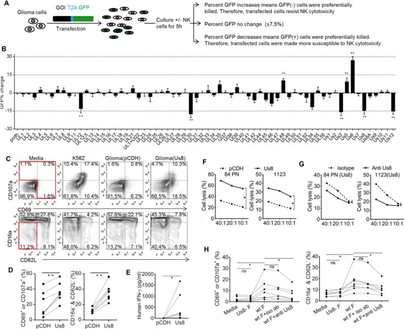
(A) Flow diagram of the DC-MEGE assay. (B) DC-MEGE results for all 65 HSV1 genes (mean ± sem, n≥4). (C) Phenotype of primary human NK cells from a representative normal donor after a 7 hour culture in media, with K562 cells (positive control), or transfected glioma cells. This was repeated with 7 individual normal donors, and a summary of the percentages of NK cells gaining the expression of CD69 or CD107a, or NK cells losing both CD16a and CD62L is provided in (D). (E) Human primary NK cells were treated as in c for 20 hours and IFNγ production was measure at 20 hours of culture by ELSIA (n=5, mean of triplicates). (F) Cytotoxicity of primary human NK cells against transfected human glioma cell lines at the specified effector: target ratio (x-axis). (G) Cytotoxicity of primary human NK cells against glioma cells expressing Us8 in the presence of isotype or mouse anti-Us8 specific antibody. (H) Summary of phenotypical changes of primary human NK cells after culturing for 7 hours in plates precoated with inactivated pure viruses. In some cases, isotype or mouse Us8-specific antibody was added into plates to block Us8 (n=5–7). Each dotted line in d, e and h links data acquired from the same donor. * p<0.05, ** p<0.01. Please see also Figure S1.