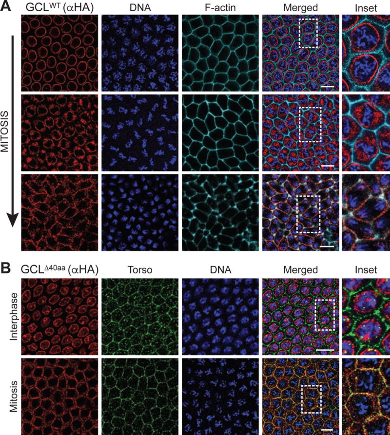
Figure 6. GCL localizes at the cortical membrane after nuclear envelope breakdown and co-localizes with Torso during mitosis.

(A) Central region of embryos of nuclear cycle 12–13 expressing FLAG-HA-tagged GCLWT. Fixed embryos were immunostained with anti-HA (red). DNA (blue) was used to determine the cell cycle stage of each embryo, and phalloidin (cyan) labels F-actin and outlines cell membrane. Dashed rectangle highlights the area shown in inset. Scale bar = 10μm.
(B) Central region of embryos of nuclear cycle 12–13 expressing FLAG-HA-tagged during interphase or mitosis. DNA (blue) was used to determine the cell cycle stage of each embryo. Fixed embryos were immunostained with anti-HA (red) and Torso (green) to assess co-localization. Dashed rectangle highlights the area shown in inset. Scale bar = 10μm.
