
Figure 1. Sirtuin Function and Localization.
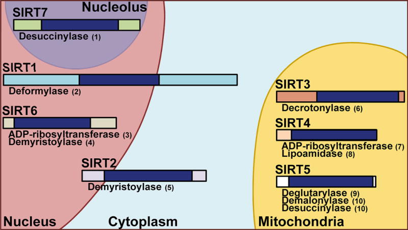
A visual representation of the seven mammalian sirtuins. Relative size and non-deacetylation activities are shown, as well as subcellular location. SIRTs 1 and 2 function in both the nuclear and cytoplasm, with SIRT1 showing more nuclear functions and SIRT2 more cytoplasmic. SIRT 6 is found in the nucleus and SIRT7 is associated with the nucleolus, while SIRTs 3–5 are primarily mitochondrial. Dark blue bands represent the conserved core catalytic domains while surrounding bands indicate the N- and C-terminal regions of each sirtuin. References for the non-deacetylation activities are as follows: (1) [87]; (2) [88]; (3) [89]; (4) [90]; (5) [91]; (6) [92]; (7) [93]; (8) [94]; (9) [95]; (10) [96].