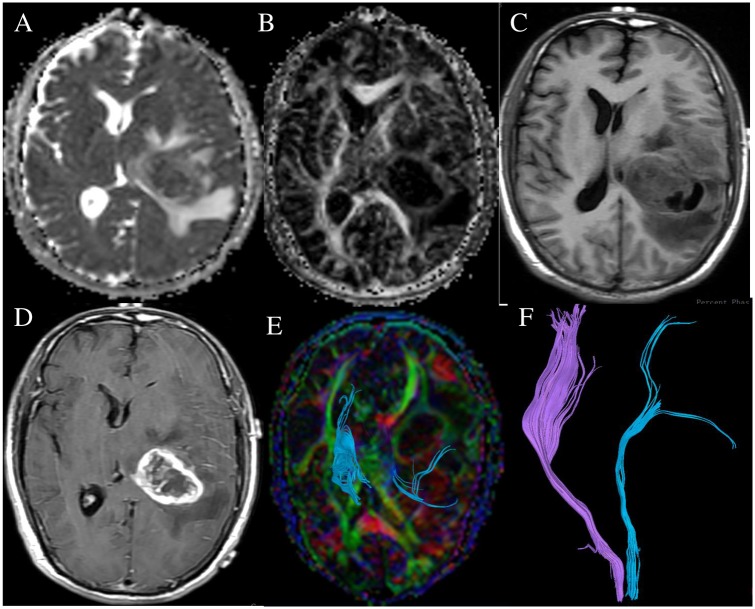
Fig 2. A 48-year-old female with left thalamic glioblastoma multiforme (WHO grade IV), the right knee muscle strength was MMT 3.
ADC map (A), FA map (B),T1WI(C), contrast-enhanced T1WI (D) show the tumor infiltrated normal tissues. DTT map (E, F) show the left corticospinal tract was displaced and infiltrated, and the number of left corticospinal fibers decreased.

