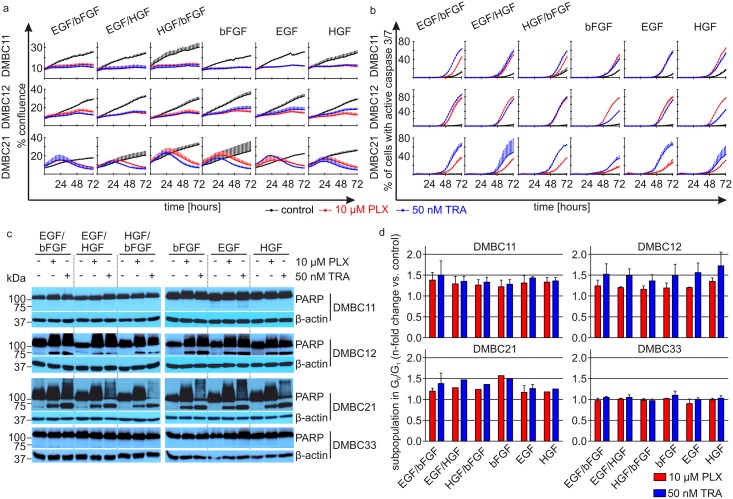
Fig 2. Composition of exogenous growth factors (bFGF, HGF and EGF) does not substantially affect vemurafenib (PLX)- and trametinib (TRA)-induced changes in melanoma cell proliferation, the induction of apoptosis and cell cycle arrest in G0/G1.
a. Proliferation time-courses. V600EBRAF melanoma cell proliferation was monitored by analyzing the occupied area (% confluence) of cell images over time using IncuCyte. Effects of 10 μM PLX and 50 nM TRA were observed in all tested populations, and reached similar levels regardless of growth conditions. b. c. Apoptosis induced by PLX and TRA in melanoma cells grown in the presence of different growth factors. b. Percent of apoptotic cells with high caspase 3/7 activity was assessed in time-lapse imaging system IncuCyte over the course of 72 h. c. Apoptosis shown as PARP cleavage after 44 h of incubation with drugs. β-actin was used as a loading control. d. Distribution of melanoma cells in cell cycle phases was determined by flow cytometry. ModFit LT 3.0 software was used to calculate the percentages of viable cells in cell-cycle phases. Drug-induced cell accumulation in G0/G1 phase was shown relatively to the respective control. Bars represent the mean values ± SD. The histograms for DMBC12 and DMBC33 cells are shown in S1 Fig.

