
Fig. 4.
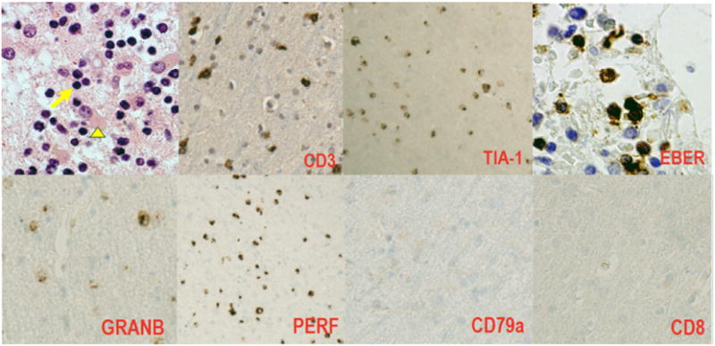
Right frontal brain biopsy: Parenchymal Infiltrate of medium sized lymphoid cells with mild to moderate cytologic atypia (arrow) with reactive astrocytosis (arrowhead). No evidence of microglial nodules, neuronophagia, vasculitis, or necrosis to suggest an encephalitic process, H&E, 40×. Immunohistochemistry demonstrates the atypical lymphoid cells are of NK derivation (CD3+, TIA-1+, EBV+, Granzyme B+ Perforin+, CD79a− CD8−).