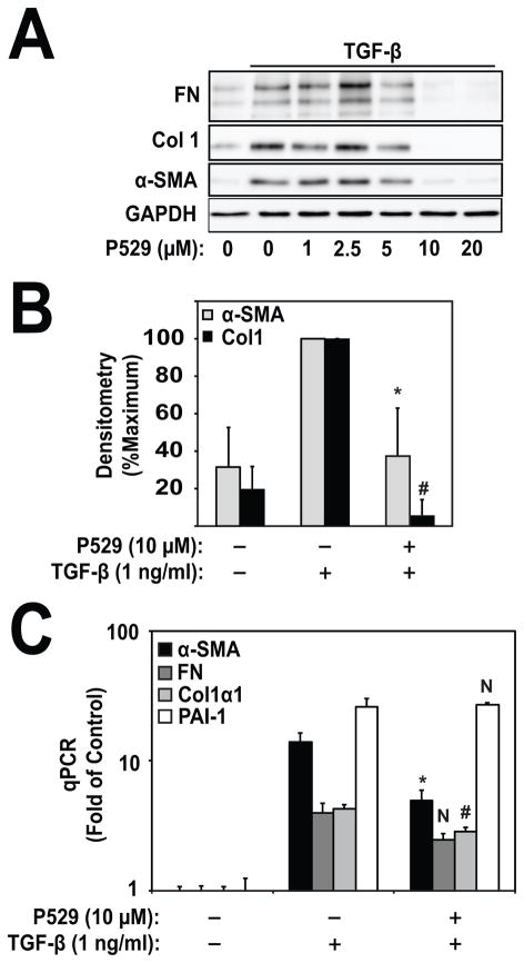
Figure 2. P529 Inhibits myofibroblast differentiation.
A) HLF were treated with 1 ng/ml TGF-β for 24 h and the indicated concentrations of P529. Lysates were subjected to western blotting for α-SMA, collagen-1, fibronectin, and GAPDH. N=3 biologic repeats. B) Densitometry of Col1/GAPDH and α-SMA/GAPDH. Measurements are normalized to the TGF-β treatment condition. Statistical significance was tested using student’s t-test. *, # denote statistically significant differences (p<0.05) between expression of α-SMA (*) or Col1 (#) in the TGF-β and TGF-β/P529 treatment conditions. C) Real time PCR of the indicated genes. Statistical significance was tested using student’s t-test. *, # denote statistically significant differences (p<0.05) between expression of α-SMA (*) or Col1 (#) in the TGF-β and TGF-β/P529 treatment conditions. N, denotes not statistically significant (FN and PAI-1).

