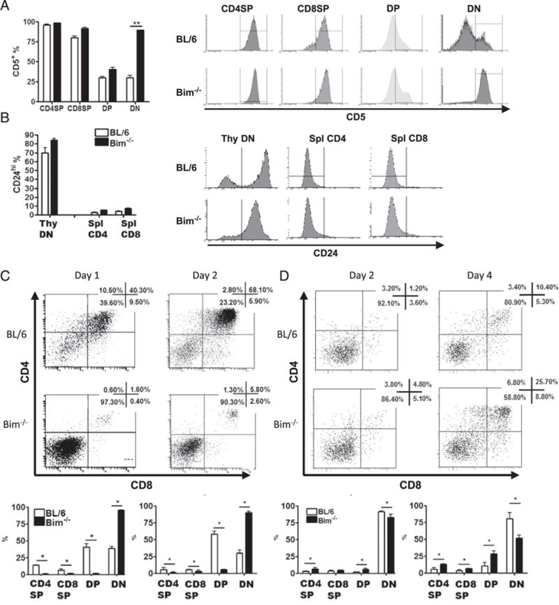
FIGURE 4.

TCR+DN thymocytes in Bim−/− mice express postselection and immature markers and do not develop into DP thymocytes in OP9-DL1 culture. Data show the percentage and histograms of thymocytes or splenic T cells expressing (A) the postselection marker CD5 or (B) the immature marker CD24, as assessed by flow cytometry. (C) Sorted DN4 (CD4−CD8−CD44−CD25−) or (D) DN1–3 (CD4−CD8−CD44+CD25−, CD4−CD8−CD44+CD25+, and CD4−CD8−CD44− CD25+) thymocytes from C57BL/6 (BL/6) or Bim−/− mice were cultured with OP9-DL1 cells and IL-7 (1 ng/ml) for indicated time and analyzed by flow cytometry. Numbers in representative dot plots show the frequency of corresponding populations. The bar graphs show the frequency of each population from either BL/6 (open bars) or Bim−/− (filled bars) mice. Results are representative of at least two independent experiments and show mean ± SD. *p < 0.05, **p < 0.01. BL/6, C57BL/6.
