
Fig. 1.
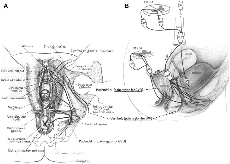
(A) Graphic illustration of female lower genital tract showing external vulva, vestibule, and lower vaginal tract. Associated innervation mediating sensory-motor activity of the pelvic region also represented. (B) Schematic drawing showing the innervation of the pelvic floor in females. Although this diagram attempts to show the innervation in humans, much of the anatomic information is derived from animal data. CEL, celiac plexus; DRG, dorsal root ganglion; HGP, hypogastric plexus; IHP, inferior hypogastric plexus; PSN, pelvic splanchnic nerve; PUD, pudendal nerve; SA, short adrenergic projections; SAC, sacral plexus; SCG, sympathetic chain ganglion; SHP, superior hypogastric plexus; Vag., vagina. (Source: Wesselmann U, Burnett AL, Heinberg LJ. The urogenital and rectal pain syndromes. PAIN® 1997;73:269-294 [62]. This figure has been reproduced with permission from the International Association for the Study of Pain (IASP). This figure may not be reproduced for any other purpose without permission.)