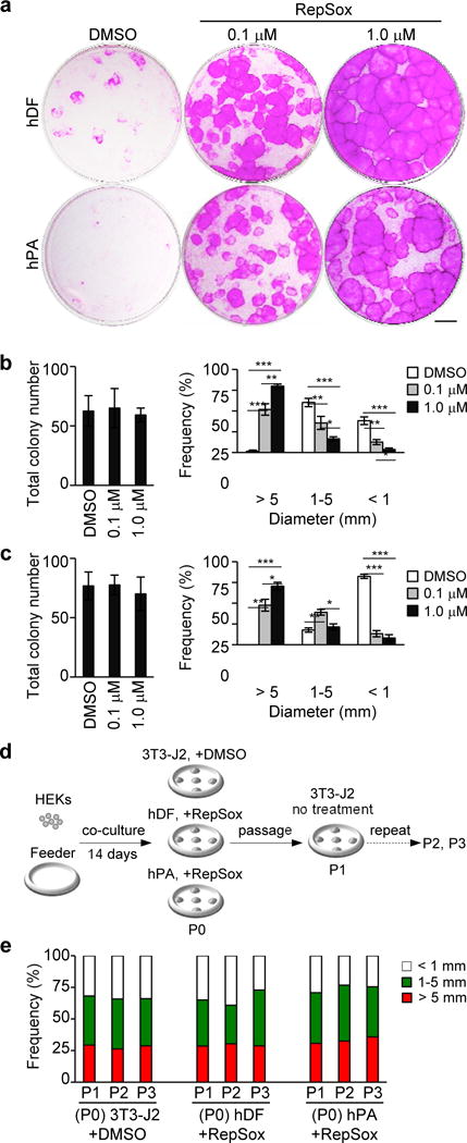
Figure 2. Inhibition of TGF-β signaling promotes the expansion of HEKs with high proliferative potential in co-culture with human feeder cells.

(a) Rhodamine B staining of epidermal cell clones grown for 14 days in co-culture with human dermal fibroblasts (hDF) or human preadipocytes (hPA) in the absence or presence of 0.1–1.0 μM RepSox as indicated. Equal numbers of HEKs (1×10³ cells) were plated in each well. Data shown are representative of three independent experiments with similar results. Bar=5 mm.
(b, c) Total number (left) and size distribution (right) of epidermal clones in co-culture with hDF (b) and hPA (c). Data shown are mean±SEM (n=3). *P<0.05; **P<0.01; ***P<0.001.
(d, e) Experimental scheme (d) and size distribution of epidermal clones in serial 3T3-J2 co-cultures (e). Epidermal cells were harvested from a standard 3T3-J2 co-culture (+DMSO) and 1 μM RepSox-treated co-cultures with hDF or hPA feeder cells (P0), followed by serial cultivation of equal numbers of epidermal cells in 3T3-J2 co-cultures with two-week intervals in the absence of RepSox (P1-P3). Data shown in (e) are representative of three independent experiments with similar results. P, passage number.
