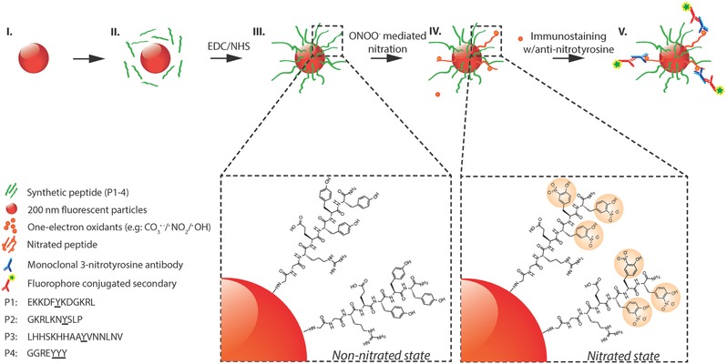
Figure 1.

Schematic representation of peroxynitrite‐induced nitration. (I) The fluorescent particle, (II) conjugation of peptides with EDC/NHS cross‐linker (P1–P4), (III) non‐nitrated peptides conjugated to surface of fluorescent particles, (IV) nitration of tyrosine through peroxynitrite‐mediated pathway, (V) immunostaining of nitrated peptides with anti‐nitrotyrosine IgGs and fluorescent secondary IgGs. (Steps I–III): Carboxyl‐functionalized red fluorescent particles (≈200 nm in size) are coated with tyrosine‐containing peptides (P1–P4, green strands). Step IV: Peroxynitrite‐mediated nitration of tyrosine residues resulting in the formation of 3‐nitrotyrosine. Step V: Immunostaining of nitrated peptides with monoclonal anti‐nitrotyrosine IgGs (MAB5404; Millipore) and fluorophore‐conjugated secondary IgGs.
