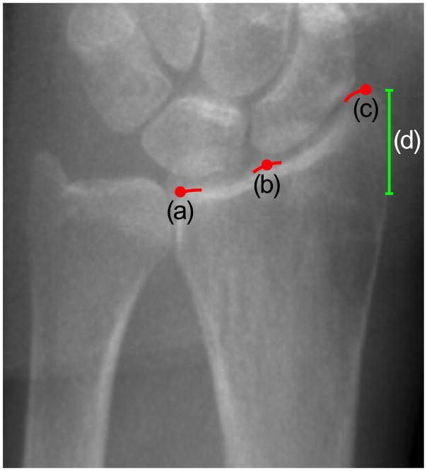
Figure 5.
Possible landmarks on the radius joint surface: (a) medial margin, (b) the notch defined by the scaphoid and lunate fossae and (c) lateral margin of the radiocarpal joint surface. Currently, HR-pQCT operators position the reference line that defines the region to be scanned at the notch between the scaphoid and lunate fossa of the radius (b). Medial (a) and lateral (c) margins represent the possible candidates for a more visible and easily identifiable anatomical landmark. The medial margin (a) might be preferable because it would exclude the subject-dependent joint height (d).

