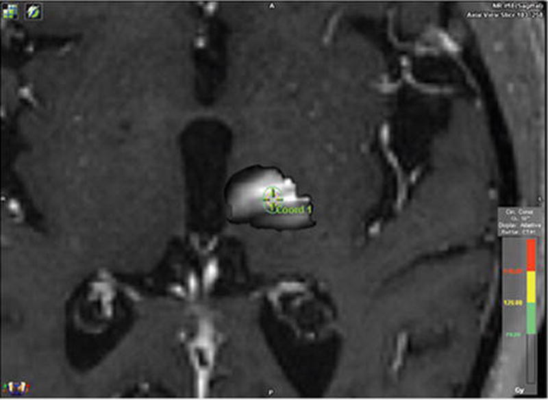
FIG. 1.

DTI-determined VIM thalamus. FSL was used to determine the areas of the thalamus with the greatest likelihood of connectivity with the premotor and supplementary motor areas. The areas of greatest likelihood for connectivity to these regions and thus the greatest likelihood of being the VIM are the whitest regions in our thalamic map. We attempted to center our isocenter as centrally as possible based on this connectivity map. Figure is available in color online only.
