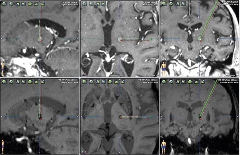
FIG. 2.

Comparison of structural MRI-determined versus DTI-determined VIM thalamic targets. The green crosshair indicates the DTI-determined VIM thalamus and shows good concordance with the ultimate radiosurgical lesion. The red dot represents the VIM thalamus target determined by standard measurements based on the AC-PC distance and third ventricle width. Figure is available in color online only.
