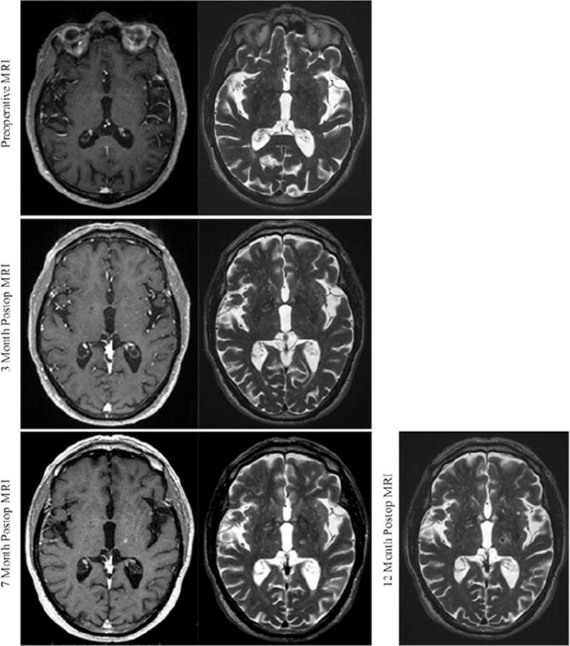
FIG. 3.

Radiographic evolution of the radiosurgical lesion. The patient was monitored radiographically, with MR images obtained preoperatively and at 3, 7, and 12 months postoperatively. T1-weighted with contrast and T2-weighted sequences were obtained at all time points except at 1 year postoperatively, when contrast scans were not obtained. At as early as 3 months, there is subtle contrast enhancement and T2 hyperintensity within the area of our intended target. These signal characteristics continue to solidify through 7 months following SRS. Twelve months postoperatively, there is T2 hypointensity within the radiosurgical target.
