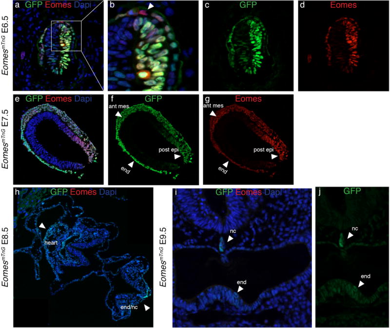
Figure 3. Comparison of endogenous EOMES protein with GFP indicates that the reporter serves as a short-term lineage tracer during gastrulation.

At E6.5 the expression of GFP (green) and EOMES (red) in the epiblast overlaps (a, c, and d), while some cells in the VE are positive for EOMES and not for GFP (arrowhead in b). At E7.5 GFP and EOMES proteins also show a gross overlap (e–g). Of note, the intensities of the stainings are different for GFP and EOMES. GFP is more strongly detected in the anterior mesoderm (ant mes) and endoderm (end) and EOMES protein shows strongest staining intensities in the posterior epiblast (post epi) (arrowheads in f and g). h) At E8.5 no EOMES protein can be detected by antibody staining while GFP protein is still present in the heart, the notochord, and the endoderm (end/nc) (arrowheads). i and j) Weak GFP expression remains detectable in some endoderm cells (end) and the notochord (nc) until E9.5, while EOMES protein is absent at this stage. All embryos are oriented with anterior to the left and posterior to the right, except for i and j where sections are oriented with dorsal to the top and ventral to the bottom.
