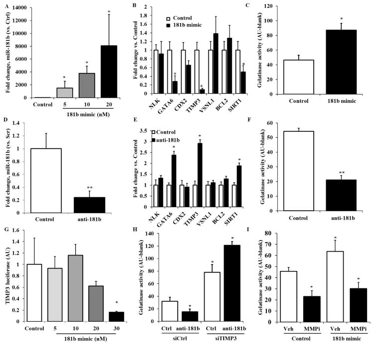
Figure 2. miR-181b targets TIMP3 and increases matrix degradation in HAVECs.
A) HAVECS were treated with miR-181b mimic at 20nM for 48h and expression of miR-181 miRNAs was quantified by qPCR. B) Expression of predicted gene targets of miR-181b was measured in HAVECs treated with miR-181b mimic or control 48h after transfection. C) MMP activity was measured via gelatinase assay in HAVECs transfected with miR-181b mimic. D) HAVECs were treated with anti-miR-181b at 100nM for 48h and expression of miR-181b was quantified by qPCR. E) Expression of predicted gene targets of miR-181b were measured in HAVECs treated with anti-miR-181b or anti-miR control 48h after transfection. F) MMP activity was measured via gelatinase assay in HAVECs transfected with anti-miR-181b G) HAVECs were transfected with wild-type TIMP3 3′-UTR firefly luciferase construct for 24h and with serial concentrations of miR-181b mimic for 24h. A dual-luciferase reporter assay was then used to measure transcription of the TIMP3 3′-UTR construct. H) HAVECs were transfected with miR-181b mimic or mimic control at 20nM and treated with MMP inhibitor GM6001 or vehicle control (DMSO) for 48h. MMP activity was measured using the gelatinase assay. I) HAVECs were first transfected with anti-miR-181b or anti-miR-control for 24h before being transfected with siRNA-TIMP3 or siRNA control at 50nM for 24h. We then measured MMP activity on these samples. n=3, *p<0.05

