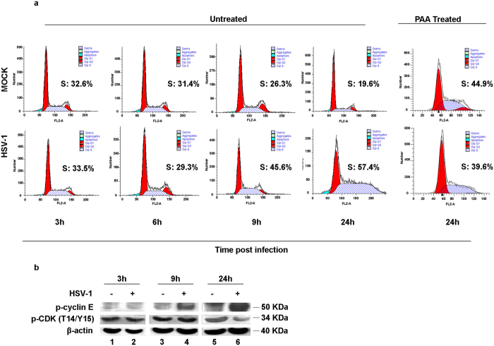
Figure 1.
Cell cycle analysis and expression of p-cyclin E and p-CDK in HEp-2 cells during HSV-1 infection. (a) HEp-2 cells were mock infected or infected with HSV-1 at MOI 10, and collected at 3, 6, 9 and 24 h p.i. At 24 h cells were infected or mock infected incubated in presence of DNA polymerase inhibitor phosphonoacetic acid (PAA). The cellular DNA content was determined at indicated times by FACS analysis as described in Methods. Processed cells were labelled with PI (10 µg/ml) to calculate different phases of cell cycle. The data were analyzed as means of triplicate ± SD and the percentage of S-phase cells was obtained using ModFit LT 3.0 software. (b) HEp-2 cells were mock infected or infected with HSV-1 at MOI 10 and collected at 3, 9 and 24 h p.i. for proteins extraction. Equal amount of proteins were separated by polyacrylamide gel electrophoresis and probed with phospho-cyclin E (Thr 395)-R and phospho-CDK (T14/Y15) antibodies. β-actin was used as a loading control.

