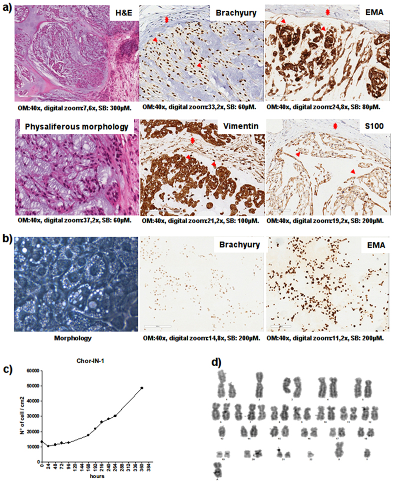
Figure 1.
Tumor and Chor-IN-1 cell line characterization by H&E and IHC. (a) The original tumor sample and (b) the derived Chor-IN-1 cell line were characterized by H&E, revealing the typical physaliferous cells, and by IHC, showing positivity for brachyury and for other chordoma typical biomarkers, as indicated. In (a), arrows and stars indicate examples of tumor and fibroblast cells, respectively. (c) Growth curve: the doubling time of Chor-IN-1 cell line was calculated as reported and found to be of about seven days. (d) Karyotype analysis of Chor-IN-1 cell line.

