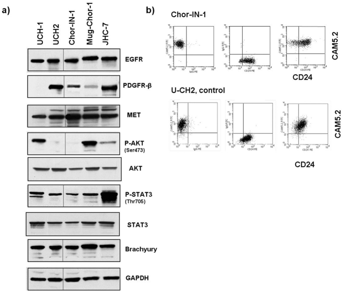
Figure 2.
Molecular Characterization of Chor-IN-1 cell line. (a) Immunoblot analysis of key tyrosine kinase receptors and other signaling molecules: Chor-IN-1, in parallel with U-CH1, U-CH2, MUG-Chor1 and JHC7 cells were seeded as indicated and collected at 70% confluence. Protein cell extracts were resolved by SDS-PAGE and filters probed with the indicated antibodies. Full-length blots are presented in Supplementary Figure 6. (b) Flow Cytometry analysis of chordoma typical membrane proteins: the expression of CD24 and CAM5.2 membrane antigens in Chor-IN-1 was confirmed to be comparable to that of the U-CH2 cell line used as reference. Upper cytograms refer to Chor-IN-1 cells and lower cytograms refer to UCH-2 cells. Left = FITC CAM5.2 vs. PE isotype ctrl: 100% of both cell lines express the marker, in the absence of non-specific signals. Middle = PE-CD24 vs. FITC isotype ctrl: more than 90% of both cell lines express the marker, in the absence of non-specific signals. Right = FITC CAM5.2 vs. PE-CD24: both cell lines are more than 90% CD24/CAM5.2 double positive, confirming both markers are expressed at levels comparable to that of UCH-2 reference cell line.

