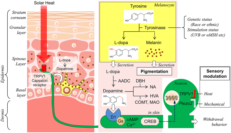
Figure 5.
Mechanism of skin dopaminergic sensory modulation. The left schema represents dopaminergic signalling (white arrow) between nerve endings (green) and melanocytes (yellow) in structure of the epithelial skin. The right schema shows tyrosinase cascade in melanocytes, metabolism of L-dopa in extracellular space (pink) in the skin and presumed CREB-dependent gene modulation pathway from nerve endings to neuronal soma of primary sensory neurons, following dopaminergic D1 receptor activation. Tyrosinase activity is different among races or ethnicity (genetic status) and environment (stimulation status). A red arrow with TRPV1 and a white arrow with Piezo2 indicate up-regulation and down regulation, respectively. TRPV1 and Piezo2 are not always co-expressed although we draw both in the same neuronal soma. AADC, Aromatic L-amino acid decarboxylase; αMSH, α-Melanocyte stimulating hormone; COMT, catechol-O-methyltransferase; DBH, dopamine-β-hydroxylase; MAO, monoamine oxidase A; HVA, homovanillic acid; UVB, ultraviolet B.

