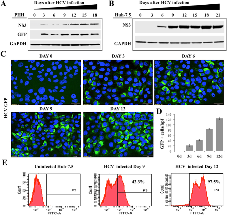
Figure 1.
Shows level of HCV replication in non-proliferative primary human hepatocytes and proliferative Huh-7.5 cell cultures. PHHs were infected with HCV-GFP virus (JFH-AM120) at a MOI of 0.1 by overnight incubation. The next day cells were incubated with fresh media with 10% human serum. (A) HCV replication in PHHs model was confirmed by the measurement of NS3 and NS5A-GFP fusion protein levels by Western blot. (B) Western blot analysis showed time dependent increase in the expression of NS3 protein in Huh-7.5 cells infected with HCV-GFP virus. (C) Expression of NS5A-GFP chimera protein with DAPI staining in Huh-7.5 cells infected with HCV-GFP virus over 12 days. (D) Quantification of NS5A-GFP expression by ImageJ software. (E) Flow cytometry analysis shows expression of GFP in infected Huh-7.5 cells at day 0, 9 and 12 days. Fluorescence images shown in panel C were taken at 40X magnification.

