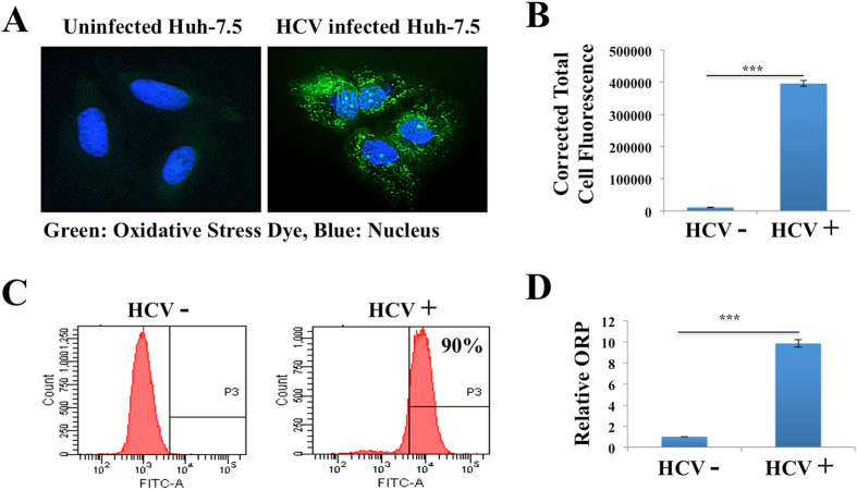
Figure 3.
Oxidative stress response in persistently HCV infected Huh-7.5 cells. (A) Oxidative stress due to generation of ROS was measured using conventional fluorescence microscopy. In this assay, the nonfluorescent dye (H2DCFDA) is oxidized to a fluorescence green (DFA) by ROS only in HCV infected cells. Uninfected cells show no fluorescence. (B) Quantification of fluorescent intensity in three different areas of uninfected and infected culture was measured by ImageJ software. Fluorescence values are proportional to intracellular ROS. (C) Flow cytometric analysis of oxidized fluorescence of uninfected and infected Huh-7.5 cells. (D) Relative Oxidation-Reduction Potential (ORP) of cell free media of uninfected and HCV infected Huh-7.5 cells.

