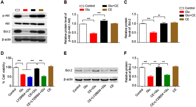
Figure 4.

Involvement of the PI3K/Akt pathway in the neuroprotective effect of CE. (A,B and C) Cell lysates were analysed by immunoblotting to measure the relative levels of proteins involved in the Akt signalling pathway, including phosphorylated (Ser473) Akt and its downstream target Bcl-2 (A). Quantification of p-Akt and Bcl-2 protein levels is shown in (B and C), respectively. (D) Cell viability was measured using the MTT assay. In this experiment, cells were pretreated with the Akt inhibitor LY29004 for 30 min prior to treatment with CE or glutamate. (E and F) Immunoblot showing the Bcl-2 protein level (E); the intensity of the bands in each lane was quantified (F). (B,C and F), n = 3; D, n = 5; data are shown as mean ± SEM; One-way ANOVA followed by Tukey’s multiple comparisons test. **p < 0.01, ***p < 0.001.
