Fig. 1.
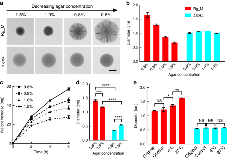
Osmotic pressure drives V. cholerae colony expansion. a Representative images of colony biofilms of the rugose (Rg_M) and EPS− (ΔvpsL) V. cholerae strains grown for 2 days on LB medium containing the designated percentages of agar. Scale bar: 0.5 cm. b Colony biofilm diameter as a function of agar concentration (rugose (Rg_M: red) and EPS− (ΔvpsL: cyan)). c To mimic the swelling effect that occurs in colonies exposed to osmotic pressure changes, 20 μL liquid droplets of LB containing 15% dextran (~2000 kDa) were placed on semipermeable membranes on top of LB medium solidified with the designated concentrations of agar. Increases in the droplet weight were measured over time. d Diameters of 4-day-old colony biofilms of Rg_M (red) and ΔvpsL (cyan) strains grown on semipermeable membranes placed on LB medium solidified with the specified percentages of agar. Unpaired t-tests with Welch’s correction were performed for statistical analyses. e To differentiate the contribution of growth from that of passive swelling during colony expansion, Rg_M (red) and ΔvpsL (cyan) colonies were initially grown for 4 days on semipermeable membranes on top of LB medium solidified by 1.5% agar at 37 °C (denoted Original). The membranes containing the colonies were subsequently floated on LB liquid medium overnight at 4 or at 37 °C. To control for growth, a separate set of plates containing membranes with colonies that had been grown for 4 days were held at 4 °C overnight (denoted Control). In all cases, the diameters of the colonies were measured after 20 h. Paired t-tests were performed for statistical analyses. NS denotes not significant; *denotes P < 0.05, **denotes P < 0.01, ***denotes P < 0.001, and ****denotes P < 0.0001. Error bars correspond to standard deviations with n = 3 in c and n = 4 in other panels

