Fig. 6.

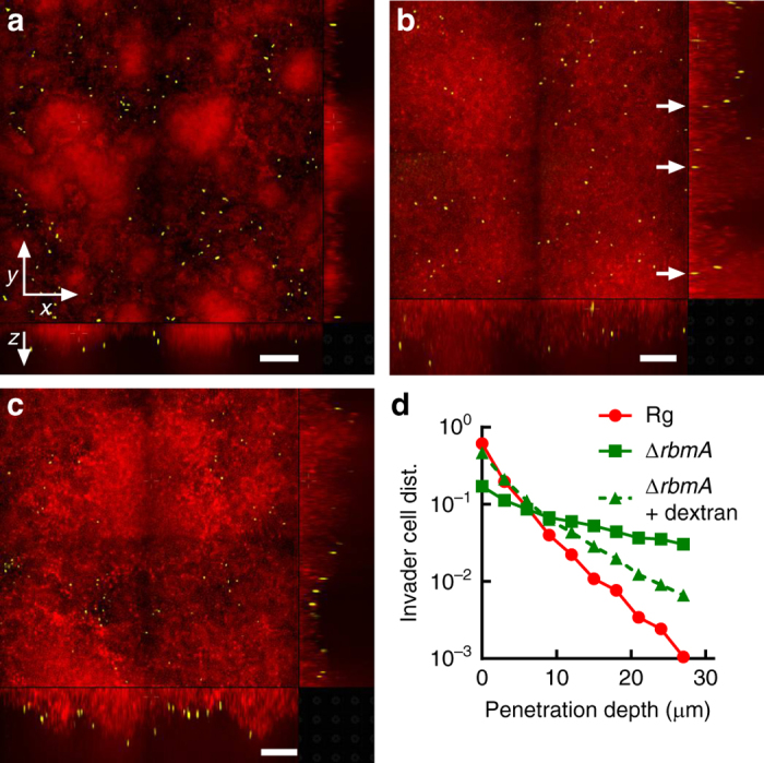
Osmotic-pressure-driven swelling modulates V. cholerae submerged biofilm susceptibility to invasion. a–c Representative cross-sectional confocal images of resident biofilms carrying mKate2 (red) invaded for 10 min by ΔvpsL strain carrying mKO (yellow). The resident biofilm strain is Rg in a and ΔrbmA in b and c. Invasions were performed in M9 medium without dextran in a and b and with 15% dextran in c. Shown are the xy cross-sectional views in the upper left panels and the two orthogonal views in the bottom and right panels. White arrows in b are guides to show invader cells that have successfully penetrated the resident ΔrbmA submerged biofilm. Scale bars: 30 μm. d Distributions (denoted dist.) of invader cells as a function of penetration depth into the resident Rg (red circles), ΔrbmA (green squares), and ΔrbmA submerged biofilms in the presence of 15% dextran (green triangles). Distributions are normalized by the total number of invader cells in each case
