
Figure 3.
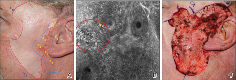
Patient with 8 prior surgeries for excision of lentigo maligna melanoma on the left cheek (A). The blue line outlines Wood lamp margins. The red line outlines the site of a prior graft. Ten mapping biopsies were performed guided by reflectance confocal microscopy. Eight were from sites with positive findings (yellow asterisks) and were confirmed histologically as lentigo maligna. Two biopsies were taken at clinically suspicious areas without positive features (blue asterisks) and showed solar lentigines on histology. Reflectance confocal microscopy showed clusters of large, round, atypical cells (red circle) with some invading hair follicles (yellow asterisk), suggestive of lentigo maligna and confirmed on biopsy (B). Other features observed included atypical pagetoid cells and dendritic processes invading the hair follicles. Final surgical defect after clinical, dermoscopic, Wood lamp, and confocal evaluation (C). Repair included removal of the prior grafts and replacement with a new split-thickness skin graft from the abdomen.