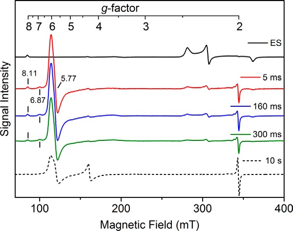
Figure 7.

EPR analysis of the enzyme–substrate complex (250 μm) reacting with peracetic acid (10 mm). The black trace shows the ES complex of cYY and CYP121. A new high-spin ferric heme intermediate is formed in 5 ms with concomitant decay of the low-spin ES complex (red trace). The EPR spectra also include the new high-spin intermediate at 160 (blue) and 300 ms (green). The final decayed reaction complex after 10 s of reaction is shown by a dashed line, which does not contain the intermediate. EPR spectra were obtained at 5 K, 9.6-GHz microwave frequency, and 1-milliwatt microwave power. mT, milliteslas.
