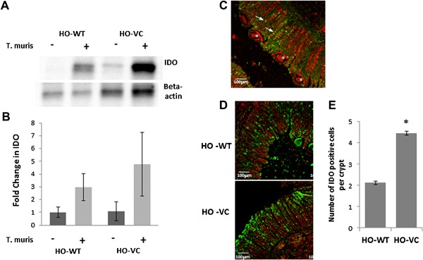
Figure 4.

Mucosal IDO protein in HO‐VC and HO‐WT cecum from uninfected and infected mice at day 35 post inoculation. (A) Representative Western blot depicting mean IDO protein in cecal mucosal scrapes normalised to beta actin. (B) Mean IDO (±SEM) in cecal mucosa as determined by Western blot. Uninfected HO‐WT n = 6, HO‐VC n = 8. Infected HO‐WT n = 11, HO‐VC n = 7. (C) Representative sections of IDO (green) and muc2 (red) stained cecum of infected mice. Arrows point to dual‐labeled cells and asterix indicate worms. Magnification‐10×, scale bar = 100 μm. (D) Representative sections of IDO (green) staining in infected HO‐WT and HO‐VC cecal tissue, propidium iodide = red. Magnification‐ 10×, scale bar = 100 μm. (n = 5). (E) Quantification of IDO positive cells per crypt in infected cecum HO‐WT n = 8, HO‐VC n = 8, ±SEM, *p = 0.01.
