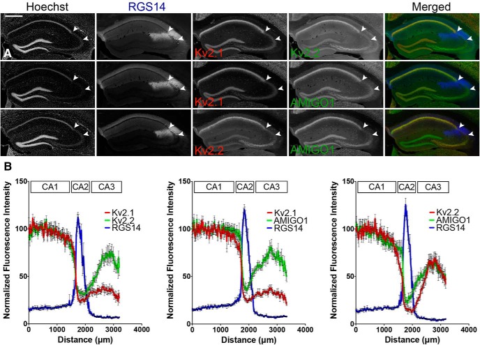
Figure 1.
The distribution of Kv2 channel α and auxiliary subunit immunolabeling in s.p. changes at the CA1-CA2 boundary, and again within region CA3 of mouse hippocampus. A, Representative low-magnification (10×, wide-field mosaic) images of C57BL/6J mouse coronal brain sections immunelabeled for combinations of Kv2.1, Kv2.2, AMIGO-1, and the CA2 marker RGS14. Arrowheads indicate the boundaries of region CA2 based on RGS14 immunolabeling. Scale bar, 500 µm. B, Quantification of mean fluorescence intensity from linescans across s.p. of regions CA1, CA2, and CA3. Values are normalized to the maximum average intensity (n = 8 mice).

