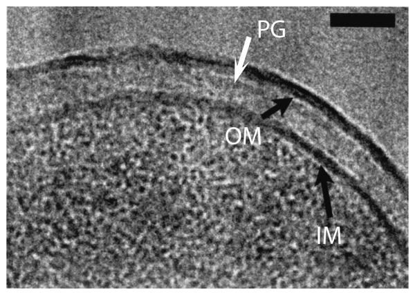
Figure 1.
CryoEM (high pressure freezing and cryosection) of the cell envelope of E. coli K-12. The black arrows point to the IM and OM. The PG is the lightly stained narrow band indicated by the white arrow. The periplasm is the lighter stained zone between the PG and IM. Bar is 50 nm. Reprinted from [16].

