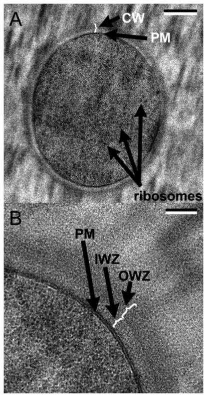
Figure 2.
CryoEM of the cell envelope of B. subtilis. A: cross section through the middle of a whole cell. B: higher magnification view. IM is designated PM (plasma membrane), periplasm is designated IWZ and the PG layer is OWZ. Bars are 200 nm (A) and 50 nm (B). Reprinted from [18].

