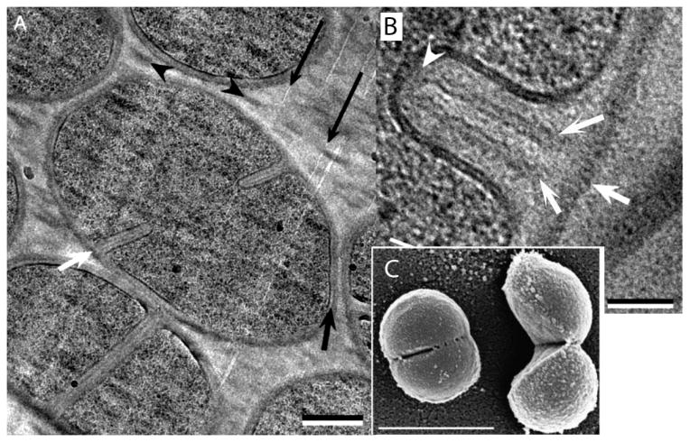
Figure 3.

Septal division of S. aureus. A: The bacterium at lower left has a completed septum but has not separated. The bacterium in the center has a septum partially invaginated. B: A newly forming septum in cross-section. White arrows indicate the PG wall of the mother cell and the two forming daughter PG layers. (C) A scanning EM of a cell just beginning to pop, and one that has just popped. (A and B) are from [33], (C) is from [34]. Bars are (A) 250 nm, (B) 50 nm, (C) 1,000 nm.
