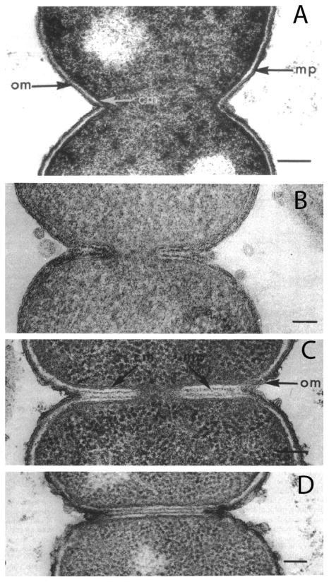
Figure 5.

E. coli division imaged by conventional embedding and sectioning following different fixation methods. A: Fixation with OsO4, the standard method in the 1970's, gives the conventional picture of constrictive division. B: Acrolein plus glutaraldehyde, a novel fixative introduced by Burdett and Murray, preserved clear septa. The two dark lines within the septa are the newly forming PG layers. C, D: E. coli CRT 97 is a chain-forming strain apparently deficient in murein hydrolases. Septa in these strains can be preserved by any fixation, OsO4 being shown here. Reprinted from [38].
